Legionnaires’ disease is of particular concern to healthcare estates managers, as it typically takes a long time to confirm the presence of Legionella bacteria in the water system and the effects of the disease can be fatal. It is estimated that up to 70% of all building water systems may have some degree of contamination,1 with both potable and non-potable systems at risk.
Potable sources include taps, showers, ice machines, humidifiers and hydrotherapy pools. Non-potable include air conditioning cooling towers; many major outbreaks have been associated with these sources.
Of the numerous species of Legionella, the primary causative species of Legionnaires’ disease is Legionella pneumophila. Between 2014 and 2016, in England and Wales there were 1,070 reported cases of Legionnaires’ diseases, with 99.7% of cases being identified as being caused by Legionella pneumophila. [Public Health England Legionnaires’ disease in residents of England and Wales – 2016. March, 2018]
For building and facilities managers, it is essential that water systems are regularly and carefully monitored and remedial actions taken as swiftly as possible when necessary.
The threat of Legionella in a water system comes from its ability to form and thrive in biofilm. Biofilm consist of colonies of initially free-swimming bacteria that secrete a protective slimy matrix to ‘glue’ themselves to inert surfaces. Biofilms build up as bacteria multiply and bind together to form a stable ‘community’.
These biofilms can develop on any damp non-sterile surface, including all surfaces in drinking water distribution networks and domestic plumbing systems.
As the biofilm forms, the microorganisms proliferate, and can spread by breaking off to reattach elsewhere in the piping, or to emerge from the system in an aerosol. It is as an aerosol that Legionella pneumophila reaches its victims, where it is inhaled into the lungs.
Aerosols may be released indoors from showers, flushed toilets and running taps, or in the wider environment from an evaporative cooling tower; the bacteria are able to travel up to three miles in contaminated water droplets.
Managing the threat
To combat the risks of bacterial contamination, facilities managers need to establish an effective water management plan, which will be unique to each location and make a commitment to regular testing for Legionella. This may be more challenging in older hospital buildings where water systems may have been adapted and altered many times during their use.
Through good water management, the threat can be minimised, even though Legionella pneumophila can survive both in acidic conditions and at relatively high temperatures of up to 50°C.
Reducing the use of flexible hoses and plastics in piping can decrease the build-up of biofilm; but it is crucial that hot water is heated to at least to 60°C and distributed at 55°C in healthcare water systems – as advised in the UK’s safe water guidance HTM 04-01 and technical guidance HSG274 – and that cold water stays cold, ideally below 20°C.

Legiolert provides a significantly faster method for detecting and quantifying Legionella pneumophila
Maintenance of systems should involve regular inspection and cleaning (when necessary) of system components and flushing regimes to keep water moving and reduce stagnation, and if possible, ‘system dead legs’ where water can accumulate should be removed. Rubber and plastic components should be avoided and biocides should also be added to water where necessary.
Water needs to be monitored regularly to check that control measures are effective. In the UK, HSG274 and HTM 04-01 set out how to achieve best practice in water management by implementing appropriate water safety plans.
Detecting Legionella pneumophila
The traditional method for measuring the presence of Legionella bacteria in water samples involves using a complex agar-plate culture test involving numerous steps, which can take up to 14 days to provide confirmed results.
The test has been in use for over 30 years, but is limited by its complexity, lack of sensitivity and accuracy.
The method uses membrane filtration to concentrate large volume samples and harvest bacterial cells. Target organisms may be lost or damaged during this process, and any non-target bacteria in the sample may overwhelm slower-growing Legionella. This can lead to results being discarded without establishing whether Legionella pneumophila may actually be present in a sample.
Samples are incubated on agar plates for 10 days using growth media containing nutrients and inhibitors to improve selectivity against non-target bacteria.
Laboratories may use various formulations from different manufacturers, and variability between lots – even from the same manufacturer – can contribute to inconsistencies between results from testing laboratories.
Results are expressed as colony-forming units (CFUs) as each observed colony is considered to represent a single bacterium from the original sample.
Since multiple plates are incubated, the result is taken from the ‘best’ plate chosen by the technician; a decision that is highly subjective as it depends on the experience and expertise of the analyst.
A secondary confirmation test is needed before final results can be delivered, lengthening the overall testing time.
Each of these sample preparation steps represents opportunities for loss, and along with the variability of methodologies, the inconsistency of competing bacteria, and the subjective nature of decision making and plate reading, increases the cumulative measurement uncertainty and the difficulty in achieving reproducible and consistent results.
Besides culture testing, recent developments in faster and more sensitive techniques such as real-time molecular techniques, provide more options for analysis, but these also have their downsides. For example, a technique such as polymerase chain reaction (PCR) testing can detect and quantify bacterial DNA with great sensitivity. However, the signals it provides (genomic units/l) cannot be interpreted readily against recognised action levels that reflect traditional plate culture and are expressed as colony forming units (CFU)/l. This can make accurate quantification of bacterial presence in a water sample difficult, particularly post-remediation efforts.

Faster culture testing
After a seven-year research and development programme, IDEXX has launched Legiolert, a new liquid culture testing technology that provides a significantly faster method for detecting and quantifying Legionella pneumophila in water samples, with results available within just seven days.
Validation studies for the method have demonstrated equal and often greater accuracy and sensitivity than traditional plate testing, providing the opportunity to identify and successfully contain and control contamination before the bacteria cause disease.
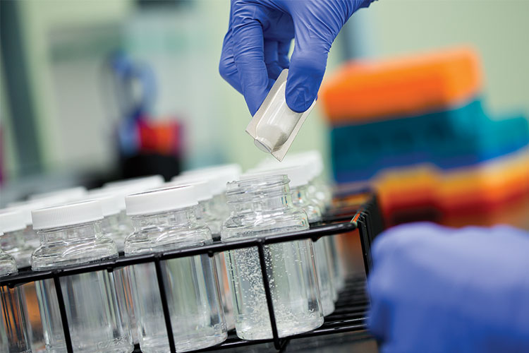
The Legiolert culture test eliminates the need for many of the complex steps described above. Instead of an agar plate, the Legiolert protocol utilises a liquid format to more closely mirror Legionella’s natural habitat. The Legiolert culture reagent is added to a 100 ml test volume; it supplies a rich source of nutrients and a suppression system to select for Legionella pneumophila. The reagent also includes a substrate indicator to provide visual detection of the target bacteria.
Ease of use
Although the protocol differs slightly for potable and non-potable water, sample preparation only takes between two and four minutes, and can be carried out by an operative with minimal training.
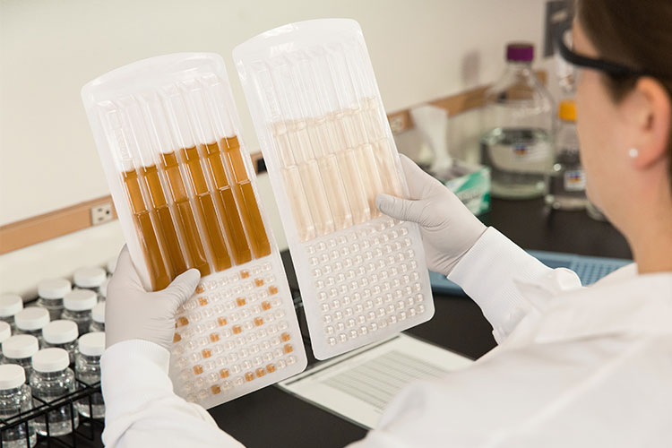
The liquid is then added to an IDEXX’s Quanti-Tray, which separates the volume into 90 small and six larger fluid reservoirs and is then sealed using a Quanti-Tray Sealer Plus device.
Legionella bacteria are slow growing, so must be incubated for at least seven days, but the highly specific colour reaction eliminates any need for further testing, making a confirmed positive result very easy to visualise in just seven days.
Following incubation, any cells in the Quanti-Tray that appear brown and/or turbid are counted as ‘positive’.
Accurate results
Subjective interpretation is eliminated and the specific nature of the indicator reduces the scope for ‘false’ negative and positive results.
The counts are made before referring to an MPN (most probable number) table to give a final result. MPN is a statistical expression of the most likely concentration in the original sample.
MPN values are equivalent to the CFU values produced in traditional testing and are an analogous measure of the action limits demanded by UK, European and US guidelines.
The Legiolert/MPN method can be used to count up to 2200, compared to the highest accurate achievable CFU count of 200 using traditional methods.
References
1. Ghose, Tia, “New York City Outbreak: What Is Legionnaire's Disease?” in Scientific American, Live Science, August 6, 2015
This article is featured in the November 2018 issue of Cleanroom Technology. The digital edition is available online.
